


Hillsdale Hospital’s Radiology Department offers state-of-the-art imaging services performed by a professional staff of technologists and interpreted by highly-trained radiologists. Highly-specialized services, such as MRI and CT, have scheduled evening and weekend appointments available. Emergency services in all areas are available 24 hours a day/seven days a week.
“Ashley in radiology is amazing! The radiologist read my report [and] I had the results before noon the same day. Everyone treated me with kindness and caring. You don’t get that personal care [elsewhere] like you do at our local hospital.” —Radiology Patient
Services Offered
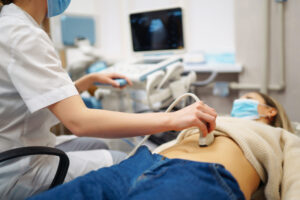
- Abdominal Ultrasound
- CT Scanner with Artificial Intelligence (AI) Positioning Technology
- DEXA Bone Mineral Density Scanning
- DEXA Body Composition Scanning
- Echocardiography
- Fluoroscopy
- Mammography with Patient-Assisted Compression Device
- MRI with Wide-Bore & Air™ Coil Technology
- Nuclear Medicine
- PET/CT Services
- Ultrasound
- X-Ray
Scheduling an Appointment
We want to make all parts of your healthcare experience readily accessible. Our dedicated radiology team makes scheduling and receiving best-in-class imaging services simple and convenient:
- Receive a referral for a radiology exam
- General X-rays are on a walk-in basis. Check in at the admitting desk on the ground floor of the hospital and our team will see you as quickly as we can.
- For other services such as MRIs or PET/CT scans, contact our radiology office. Most appointments can be scheduled within one week or less
State-of-the-Art Imaging Services
 Assess and identify bone, muscle and fat in the body, including visceral fat. This information assists you and your providers in understanding risk factors for various conditions, as well as targeting fitness and nutrition goals based on body composition.
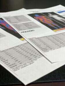
What the Assessment Shows
- Digital body composition image showing three types of tissue: bone, muscle & fat
- X-Ray with markers placed for seven body regions
- Total body fat percentage
- Body fat percentage compared to people of the same age, race and gender
- Fat Mass Index (best measure of excess fat)
- Estimate of visceral fat around organs
Why Assess Body Composition?
- Analysis of fat type and location. Certain fat types are an indicator of increased risk of cardiopulmonary disease, cancer, arthritis, diabetes, etc. Even those who have a low body weight can carry fat in areas of risk.
- Monitoring of healthy changes. Patients may use it to monitor progress of exercise and nutrition changes.
- Immediate results. Get a printed report minutes after your test. Use that report to follow up with your physician, fitness coach, dietitian or nutritional advisor for ways to improve your health.
- No insurance hassle. This service is not billable to insurance, so we offer this service at an affordable flat rate. For current pricing, call the scheduling number below.
Cross-Functional Benefits
- Dietitian: Athlete nutrition plans
- Strength & Conditioning Coach: Athlete conditioning plans
- Athletic Training & Sports Medicine: Injury prevention, rehabilitation planning
- Coaches: Realistic weight goals
- Athlete: Info to help improve performance & motivation
Get 25% off your second scan if you return within a year of your first scan!
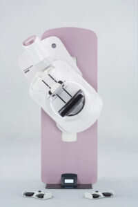
Photo courtesy of GE
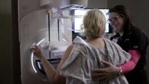
Photo courtesy of GE
A mammography system designed by women, for women. Our mammography system reduces patient anxiety and discomfort, while producing the highest-quality images with the lowest radiation exposure. The Senographe Pristina 3D mammography system from GE Healthcare allows for greater accommodation and gives patients more control during their experience. Our nurse navigator ensures that each patient has a partner through their mammography journey.
Why focus on patient comfort?
A mammogram can be challenging both mentally and physically. This system significantly minimizes discomfort and creates a better experience for our patients. Hillsdale Hospital’s new system and remodeled mammography suite have been re-designed around the patient with comfort-enhancing features, flexible positioning and sharp pictures. A new self-controlled, hand-held device allows clients to control their own breast tissue compression. The new shape and ease of the machine give us the ability to better accommodate differing shapes, sizes, heights, and mobility levels.
The Senographe Pristina 3D has been shown to bring anxiety down for 97 percent of patients.
What is 3D Mammography?
3D mammography, the next step beyond 2D, is the highest standard of care. Our 3D system captures high-quality images while automatically reducing radiation exposure to the minimum necessary.
Why have a nurse navigator?
Our nurse navigator makes the mammography process more individualized. Utilizing a nurse navigator and skilled mammography technologists, Hillsdale Hospital’s team approach with state-of-the-art technology gives patients the most up-to-date care available, while offering the personal touch that comes with locally-based healthcare. Our nurse navigator guides patients through the mammogram experience—before their scheduled appointment, during the procedure and after results are known. Our process is fast, patient-centered and thorough.
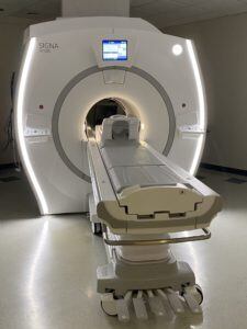
State-of-the Art Technology Built with Patient Comfort Features
If you struggle during MRI scans because your position makes you uncomfortable, you have claustrophobia, or the loud sounds of the MRI make you anxious, the new MRI tech will make your scan a more relaxed experience. Our best-in-class MRI system includes more patient comfort measures, such as a wireless headset for music during your scan, noise reduction technology, and head first or feet first entrance into the scanner—your choice. Now getting an MRI can be more comfortable and less stressful.
Hillsdale Hospital was the first public hospital in the state of Michigan to use Air™ Coil Technology on a 1.5 Tesla magnet, the standard MRI strength for hospital care.
What is Air™ Coil Technology?
The Air™ Coil is like a camera used on dedicated body parts during an MRI scan. The new technology allows the camera to be flexible and feel more like a blanket laying over the patient instead of a rigid piece of equipment. The flexibility of the coil allows improved positioning for maximum patient comfort and the ability to scan larger areas of the body at one time. This technology provides improved image quality and is able to perform two new exams we could not previously offer: MR elastography, which is used to diagnose liver stiffness caused by disease, and prostate imaging, used to identify conditions like prostate cancer.
Promote earlier diagnosis and treatment of health conditions thanks to local access to PET/CT services. PET/CT services, used to help diagnose, monitor or treat conditions like cancer, heart disease and brain disorders, are available on our campus every Wednesday, decreasing patient travel time and increasing availability of service.
How quickly can I get in?
Many PET/CT services book out weeks in advance, but we can typically get patients within seven days. This is critically important for patients who need it—especially those who require routine or frequent scans.
What is the quality of this PET/CT system?
The mobile Siemens Biograph MCT-20 provides patients with the same level of imaging technology offered at large hospitals and academic health systems. This next-generation scanner offers short scan times and a wide bore for patient comfort, as well as excellent image quality so physicians can visualize small lesions. The low-dose capability of our PET/CT system supports our efforts to maintain high levels of patient safety, while still delivering the image quality physicians need to properly diagnose, monitor and treat their patients’ conditions.
How are PET/CT scan images used?
The high image resolution and quality allows for early identification of cancerous tissues and state-of-the-art tumor staging. This supports a physician’s efforts to determine whether a treatment is working and monitor progress. As a result, physicians can limit side effects patients experience from treatments that are not working by changing or discontinuing them.
